What is Crystallography?
Scientists have several sophisticated analytical techniques at their disposal to elucidate the physical structure and properties of substances. A technology commonly employed by analytical scientists is crystallography, of which there are many variations. This article will discuss this subject.
 Image Credit: Gregory A. Pozhvanov/Shutterstock.com
Crystallography – An Overview
Crystallography is the term applied to the field which studies crystalline structures. Many crystalline structures exist in nature, such as gemstones and rocks. In the field of life sciences, however, crystallography is primarily used to study proteins and other important biological molecules which do not crystallize naturally.
Before the advent of crystallography, the study of crystals was based upon physical measurements of their geometry to establish their symmetry. The field originated with the discovery of X-Rays in 1895. In the early 20th century, pioneering work was carried out by H. and W.L Bragg, who demonstrated the relationship between crystalline structure and X-ray diffraction patterns. This formula is known as Bragg’s law.
The field of crystallography has revolutionized our understanding of the structure of minerals, proteins, DNA, viruses, and pharmaceutical materials, to name but a few examples. Over the course of the past century, the field has evolved, with many crystallographic techniques developed.
Principles and Theory
Crystallography methods analyze diffraction patterns of a substance caused by shining a beam of radiation at it. Electromagnetic radiation such as X-rays are the most common, but neutrons and electrons are also used. The three types of beams – X-ray, electron, or neutron – interact with the substance in different ways. Each is suited to different studies.
The diffraction patterns created by X-rays are due to interaction with valance electrons and their spatial distribution. Electron beams detect the charge distribution in atomic nuclei and their surrounding electrons. Neutron beams undergo scattering by forces such as strong nuclear forces and magnetic fields.
X-Ray Crystallography in Pharmaceutical Drug Development
Crystallography is used to study atomic structures because optical microscopy is unsuitable for this purpose. This is because visible light’s wavelength is several magnitudes larger than the length of atomic bonds and atoms. The shorter wavelength of electromagnetic radiation is, therefore, more suited to the analysis of structures at this scale.
A key difference between crystallography and optical techniques is that the beam cannot be focused to produce images as such. Rather, the structure is reconstructed from the diffraction pattern using a variety of techniques including software.
In the diffraction pattern produced by crystallographic techniques, sharp features are caused by photons reflected by the atoms in the structure, whereas diffuse and weak features are caused by non-periodic components. Due to the repetitive and ordered nature of crystal lattices, diffraction patterns created by crystallographic methods are ideal for providing information on the structure of solid materials.
Technique
As has been mentioned, the three types of diffusion techniques used in crystallography are X-rays, neutrons, and electrons. Many other analytical techniques are also employed in crystallographic studies, such as X-ray fluorescence, spectroscopic techniques, and computer visualization and modeling.
First, a stable crystal must be created. As many biological molecules do not occur naturally as crystals, crystallographers employ a variety of techniques. Some substances crystallize more easily than others.
To form the crystal, the molecule is held in a solution, and along with a buffer and precipitants is sealed within a reservoir. This reservoir contains a hygroscopic solution, Crystallization is achieved by the steady precipitation of the solution containing the molecule into the reservoir. The process is a slow one – if it is too fast, a proper crystal cannot be formed.
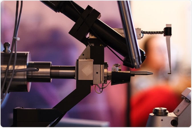
Once a stable crystal has been formed, it is introduced to the crystallographic equipment. Then, a beam is directed at it. Synchrotrons are the most common source for the beam due to the purity of emitted radiation. They also produce the most complete diffraction patterns. Other sources exist, but synchrotrons remain the standard
To analyze the produced diffraction patterns, incredibly complex mathematics are used. Two generated pattern is compared to a pattern which has been predicted mathematically. The process goes through several refinements before the true pattern of the crystal is determined. Modern software has greatly reduced the need for revisions. Without software, the process of elucidating the crystals form can be incredibly laborious and time-consuming.
Whilst less ordered, patterns can be formed by powders and fibers. The diffraction pattern created by these types of substance can still provide useful information due to an inherent degree of order. One example of this is the discovery of the structure of DNA, where the diffraction pattern which revealed its double-helical structure was obtained from a fiber sample.
In Conclusion
Crystallography is a robust analytical technique that has been used in a variety of fields such as life sciences, geology, and materials science. It has been used to reveal the structure of viruses, proteins, nucleic acids, gemstones, and industrially important substances. It remains one of the gold standards of analytical chemistry.
References:
- BCA Education (Website) History of Crystallography [Accessed online 13th July 2021] https://learn.crystallography.org.uk/learn-crystallography/history
- New World Encyclopedia (Website) Crystallography [Accessed online 13th July 2021] https://www.newworldencyclopedia.org/entry/Crystallography
- Chemeurope (Website) Crystallography [Accessed online 13th July 2021] https://www.chemeurope.com/en/encyclopedia/Crystallography.html
Further Reading
- All Crystallography Content
- Protein Crystallization
- X-Ray Crystallography
- Protein Structure Determination
- Microseeding Explained
Last Updated: Oct 4, 2021

Written by
Reginald Davey
Reg Davey is a freelance copywriter and editor based in Nottingham in the United Kingdom. Writing for News Medical represents the coming together of various interests and fields he has been interested and involved in over the years, including Microbiology, Biomedical Sciences, and Environmental Science.
Source: Read Full Article
