
The critical role of flow cytometry in CAR+ T-Cell trials
 insights from industryAns De Beuckelaer & Rowan ClayesCerba Research
Please could you both introduce yourselves and give a brief outline of your roles at Cerba Research?
ADB: My name is Ans De Beuckelaer. I have a Ph.D. in biotechnology from the University of Ghent. In 2018, I joined Cerba Research as a scientist in the Flow Cytometry department. I became the EU Regional Head of the Flow team after three years in the role, and in this position I became part of many global clinical trials in respect of the development, validation, and implementation of multiple flow assays.
RC: I’m Rowan Claeys. After graduating as a medical doctor and clinical pathologist from the University of Louvain, I had roles in various hospital laboratories. In these roles, I was head of medical validation in laboratory hematology. I joined the Flow Cytometry department as a clinical pathologist at Cerba Research in 2020.
Could you tell us about the importance of flow cytometry in CAR+ T clinical research?
ADB: Being one of the leading technologies for cellular analysis, flow cytometry generates simultaneous high throughput enumeration and individual cell characterization data.
With the breakthrough of cellular immune therapies, such as CAR+ T, flow cytometry became a critical platform, not only for the clinical laboratories, but also for drug developers and manufacturers. Indeed, flow cytometry plays a crucial role in the production process of CAR+ T cells where it is used for assessing transduction efficiency, purity and characterization of the CAR+ T product before infusion. But also after manufacturing, when CAR+ T cells are infused into the patients, flow is used in clinical laboratories to assess CAR+ T expansion, efficiency and persistency and to monitor minimal residual disease (MRD).
What are the types of flow cytometry assays in CAR+ T trials?
ADB: Flow can be used to monitor on one hand CAR+ T cells and endogenous immune cells, and on the other hand circulating malignant cells.
To monitor CAR+ T cells, we run two types of flow assays: cellular pharmacokinetic (PK) assays and characterization assays. A CAR+ T PK assay is designed to enumerate CAR+ T-cells. Initially, qPCR was used for PK testing. Though, since standardization of flow testing over the globe is made easier with current cytometers, and high sensitivity can be reached using high-affinity anti-CAR reagent, flow can now be used for global PK testing in clinical trials. Exploratory CAR+ T characterization assays are designed to phenotype and assess the characteristics of both endogenous cells as well as CAR+ T cells, to gain a better understanding of the mechanism of the drug.Further, Minual Residual Disease (MDR) assay can be performed to determine the efficiency of the treatment by analyzing the liquid tumor clearance. The MRD assay can be extended by a phenotyping assay to define the presence of specific biomarkers on malignant cells, or to define antigen loss throughouth the treatment.
Please talk us through CAR+ T characterization assays.
ADB: A well-designed characterization assay gives the drug developers insight in how the CAR+ T cells behave post-infusion and how they impact the patient's endogenous immunity. The assay composition depends on the cell types and biomarkers the investigator want to explore.
CAR+ T characterization assays developed at Cerba Research are focussing mainly on the memory differentiation and activation and/or exhaustion status of expanded CAR+ T cells and endogenous immune cells. Further, they are designed to pick up non-T CAR+ cells in patients and to monitor the presence of undesired target expression on the CAR+ T surface.
What about having absolute counts reported from flowcytometry assays?
ADB: This is possible. Flow cytometry allows reporting of relative counts and absolute counts of fresh specimens. There are two main approaches: either the single platform or the dual platform. According to the single platform, the absolute counts are calculated based on commercial quantification beads or the capability of volumetric acquisition. There is a range of benefits to this approach. However, due to the presence of beads in the tubes, this technique cannot be combined with a wash step after staining, and volumetric analysis is not feasible on every instrument. Therefore, the dual platform might be considered for complex multi-color flow assays. The dual platform combines absolute counts of parent populations defined by a hematology analyzer, preferably collected in the same specimen collection tube. Absolute counts of the population of interest is then calculated by the relative percentages obtained by the flow assay and the data from the hemato analyzer. However, it should be kept in mind that these absolute counts are only semi-quantitative and should always be interpreted as an approximation. Indeed, whereas Sysmex has direct acquisition without wash steps or viability distinction, the ratio of populations within the leukocyte population might differ between the two platforms.
Therefore, as a solution for complex assays we recommend obtaining the absolute count of a parent population from a companion tube with quantification beads. In this tube, the sample is stained with a basic panel in a Lyse/No Wash format.
What are the key aspects to keep in mind when developing a CAR+ T assay?
ADB: A CAR+ T assay is well designed when it is able to report an accurate set of data for its intended use. The intended use, and regulatory requirements define the validation parameters of the assay, which is based on a fit-for-purpose principle.
Having defined the intended use, the next pillar of CAR+ assay development is the choice of the specimen type. This can be whole blood, bone marrow aspirate or PBMC. The choice is dependent on a range of factors, including the feasibility of local acquisition, the need to run in batches, the need for absolute counts, the presence of your population of interest and the stability of the parameters. PBMC is preferable in instances when stability does not allow global shipment of fresh material, or in case when batch testing is required. Further, PBMC preps enable enrichment which might be needed to monitor CAR+ Ts in heavily lympho depleted patients, which is seen in allogeneic CAR+ T therapy.
Another crucial aspect of any assay development is the selection of reagents. More specifically for CAR+ T PK assays, the performance of the anti-CAR detection antibody is crucial to reach high sensitivity. Given that anti-idiotype monoclonal antibodies have a high affinity for CAR+ T and exhibit a low nonspecific binding, they are highly recommended. A major drawback is that these reagents require customized development. The combination of a long manufacturing period, risk for interlot variability and unpredictable elements within the sample forecast makes it challenging to manage reagent stock in global trials. Therefore, commercial antibodies, such as target fusion proteins or anti-tag monoclonal antibodies are operationally seen as an easier alternative.
Another aspect to keep in mind when designing the gating strategy is the character of the CAR+T product itself. There are two types, autologous and allogeneic CAR+ T therapies. The autologous CAR+ T-cells are taken from the patient, then transduced and infused into the same patient. Therefore, gating can go with CD3 positive T-cell selection. Whereas allogeneic CAR+ T-cells are genetically knocked out for the CD3 T-cell receptor to reduce graft versus host risks, and thus require another gating strategy.
Further, but not least, it is vital to avoid risks of interference of antibody-based therapies targetting markers of interest of the assay. Examples can be anti-CD38 or PD1/PDL1 inhibitors. Clones of reagents used in the assays are to be selected based on their compatibility with the treatment of the patients.

Image credit: Cerba Research
How has this assay development and validation been approached in Cerba Research’s laboratories?
ADB: We offer customized panel design and fit-for-purpose validation, which can be fine-tuned with patient samples. Our approach is truly a scientific collaboration between Cerba Research and the sponsor with open communication, and complete transparency on validation data, SOPs and reports. Upon initial validation in EU or US, the assay is transferred and implemented around the globe, according to the need of the trial.
Cerba Research has flow cytometry capability around the globe, operating in Europe, Africa, Australia, US, Taiwan and China. We have access to 12C BD Lyric platform on almost all sites (Australia, Europe, US, Asia), allowing the organization to offer global transparency. Besides instrumental standardization, we work with global standardized validation procedures, acquisition and analysis templates and SOPs. Besides the 12C Lyric instruments, we offer flow on other platforms as well, such as +40 multicolor flow on Cytek Aurora in EU and US.
Study sample data analysis is centralized in order to guarantee alignment within the analysis team. Turnaround time is agreed upon upfront and data transfer is automated via our own data platform.
Could you provide us with an overview of the added value of flow in multiple myeloma, Minimal Residual Disease (MRD) assessments, and multiple myeloma phenotype?
RC: When it comes to immunophenotyping of the plasma cells themselves in CAR+ T-cell clinical trials, there is a great deal of added value in this approach. This is undertaken with the use of next-generation flow (NGF) cytometry that is based on the standardization of every step of the process from sample preparation over data acquisition to reporting, on the use of a selection of (more) backbone markers and reaching a higher sensitivity than 1st generation flow cytometry
At diagnosis, or as part of the screening, next-generation flow cytometry is done with the start of the CAR+ T-cell therapy in order to identify and quantify the malignant plasma cells in the bone marrow, and look at the antigen’s presence.
CAR+ T-cell therapy or other immunotherapeutic drugs will be used to target antigens on the plasma cell surface. During the treatment follow-up, NGF is also important, as targeted therapy might induce phenotypical changes over the plasma cells, and change the expression of the targetable antigens.
The disappearance of the monoclonal plasma cells proves the efficacy of the therapy. NGF can also be used to assess MRD – something which is becoming increasingly critical as a form of marker for progression-free survival and overall survival in clinical trials.
How is immunophenotyping of plasma cells in multiple myeloma performed?
RC: In multiple myeloma, immunophenotyping of plasma cells is performed with a set of recommended markers. Based on the expression of CD45, CD38 and the more plasma cell-specific marker CD138, the plasma cell populations are identified – which classifies them as normal or abnormal plasma cells.
Next, the expression of a set of other plasma cell characterizing markers is used. These malignant plasma cells tend to be either CD19 negative and CD56 positive, and additionally tend to have a weaker or absent expression of CD27 and CD81. It is only within a small part of MM patients that CD117 expression on malignant plasma cells is observed.
Finally, intracellular staining is used to assess the monoclonality of the malignant plasma cells for kappa and lambda light chains. In an ideal situation, this should lead to good discrimination of both normal and abnormal plasma cells.
There are a number of other interesting markers that could be included in multiple myeloma panels in addition to the recommended markers. These might be markers that are predictive of disease progression, such as CD28 and CD200.
It is known that CD138 is a relatively unstable marker, and its expression diminishes between 8 and 24 hours after the sample has been taken. When you know that your analysis will be delayed, it might be interesting to add a more stable marker like CD319 to the panel.
How are patients that undergo CAR+ T-cell therapy treated?
RC: The majority of patients who undergo CAR+ T-cell therapy treatment will be pre-treated with daratumumab (anti-CD38), which has an effect on the expression of CD38. As we have stated, alternative markers like CD229 and CD319 could be introduced into a panel. Therefore, it might be useful to investigate whether your targeted antigen is expressed on the malignant plasma cells before starting the CAR+ T-cell treatment or another immunotherapeutic treatment. There is a range of other markers of other antigens currently being investigated for their potential to develop a CAR+ T-cell therapy – which includes CD229, CD44, Lewis and other antigens. A number of these antigens are now under investigation and in clinical trials.
What is the impact of immunotherapeutic drugs on the phenotype of the plasma cells for current treatments?
RC: I’ll briefly outline the impact on two popular treatments: the first, daratumumab, and the second, BCMA-targeted CAR+ T-cells. The drug’s next-generation flow by CD38 receptor occupancy can be directly interfered with by treatment with daratumumab – which leads to the disappearance of the CD38 expression on the plasma cells. A special CD38 multi-epitope antibody that binds to a different epitope than daratumumab – can be used to overcome this, as can the use of an intra cytoplast staining for CD38 that is bound to the endoplasmatic reticulum. The use of other plasma cell-defining antibodies like CD229 and CD319 is also a possibility.
CD38 antigen loss is another effect of treatment with daratumumab, which may either be due to a transient phenomenon or a temporary downregulation of the CD38 antigen on the plasma cells. This can last up to six months after the last infusion of the drug.
It is important to note that low or lost CD38 expression can also be a result of the genetic selection of CD38 negative plasma cell clones in order to escape the daratumumab therapy. CD38 might also get lost due to trogocytosis by monocytes and granulocytes, as the cells can eat away at parts of the plasma cells and other antigens in the CD38 complex neighborhood. For CD56 and for CD44, this has been described. Another plasma cell-defining antibody must be used to retrieve the plasma cells: the most popular antigens here are CD229 and CD319.
What is the most frequently targeted antigen for CAR+ T-cell therapy?
RC: B cell maturation, antigen, or BCMA or CD269 is currently the most frequently targeted antigen for CAR+ T-cell therapy. In multiple myeloma patients and other patients’ plasma cells, the antigen is expressed at very high levels. It is not present in B cell precursor cells. Gamma-secretase removes the BCMA from the plasma cells and these can then be retrieved in the serum of the patients as soluble BCMA.
BCMA expression is correlated positively with disease progression. In patients with MGUS, it is low, and in patients with multiple myeloma it is higher. It is likely that there will be a reversible down-regulation of BCMA on the plasma cells after treatment with anti-BCMA CAR+ T-cells. There may be a clonal selection of BCMA negative or BCMA low expressing plasma cells as an escape mechanism, which expresses multiple myeloma cells that will proliferate.
Due to molecular aberration of chromosome 16, there may also be BCMA antigen loss. This antigen loss may also be caused by removal of BCMA from the plasma cell surface by gamma-secretase. All these phenomena will lead to relapse with BCMA, weak positive or BCMA negative plasma cells.
We may also see trogocytosis, but this time the trogocytosis will be done by the CAR+ T-cells, which will lead to an expression of BCMA on the CAR+ T-cells. Other CAR+ T-cells will then kill the CAR+ T-cells, in a phenomenon termed ‘fratricide.’
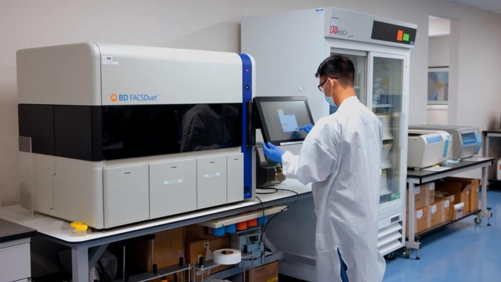
Image credit: Cerba Research
How can Next Generation Flow Cytometry (NGF) be used to look for rare plasma cells in patient bone marrow?
RC: In order to search for bone marrow of the treated patients, NGF can be used. Criteria for the response in multiple myeloma patients has been described by the international myeloma working group. This allows us to note that over 50% of the treated multiple myeloma patients will reach a complete response, which has been outlined as a presence of under 5% plasma cells in the bone marrow, the disappearance of soft tissue plasmacytoma, and negative unification in serum and urine.
It is an unfortunate truth that most of these patients will relapse – even patients in stringent complete remission, and in order to detect the persistent disease below the levels of complete remission, high sensitivity methods are needed. The IMWG has therefore added MRD negativity by flow cytometric, molecular or imaging techniques, on top of the complete remission criteria.
How else does MRD relate to clinical outcomes?
RC: When it comes to clinical outcomes – defined as progression-free survival and overall survival – MRD is also the most relevant predictor of these. It is independent of the disease stage at diagnosis, the risk profile of the patient, and the effect of whether or not they have received an autologous stem cell transplantation. At this time, therapeutic strategy is not changed depending on the patient’s MRD status, but there are many ongoing clinical trials which both investigate the optimal time for autologous stem cell transplantation and the type of consolidation therapy and its duration.
MRD is a strong predictive factor of both overall survival and progression-free survival. Regarding the accelerated release of newly developed drugs, obtaining an MRD negativity could also be used as a surrogate endpoint. However, until this point, it has been considered unacceptable for both the FDA and EMA, but negotiation may be possible for the different agencies.
In multicenter clinical trials, MRD assessment is also gaining importance where FDA and EMA have been approved. These are used as a surrogate marker for progression-free survival.
Are there any issues with the use of the assessment of MRD?
RC: Of course, it would be false to say that there are no outstanding issues that require resolution. For instance, the FDA recommends the use of MRD assessment only in patients with complete and stringent complete response, where the EMA would also include patients with a very good partial response. There have not been good descriptions for either the timing points for assessment of MRD, or the duration of the MRD monitoring.
However, it does appear likely that both agencies will agree on a period of a minimum of one year by implementing the IMWG criteria of sustained one-year MRD negativity. The two agencies seem to have agreed to employ a cutoff of 10-5. In order to exclude the presence of extramedullary disease, other methods like imaging techniques are necessary. The use of liquid biopsies could also be a possibility, but the results from NGF have not been very positive – until now. The determination of cell-free DNA promises to be a more effective technique.
Moreover, the organization also needs to perform further risk stratification for the patients that are proven to be MRD positive when clinical trials are performed. Currently, there are only two techniques that have sufficient sensitivity to assess MRD, NGF and next-generation sequencing (NGS), and both of these have their own advantages and disadvantages.
How does NGS compare to NGF?
RC: Delayed analysis is possible with NGS, thanks to the fact that no fresh sample is needed. However, it must be noted that NGF is possible in the absence of a diagnostic sample, which NGS is not. The presence of other cells over plasma cells is an indicator of both the sample quality and the possible hemodilution of the sample.
Both techniques offer their users complementary information where molecular characterization by NGS provides information on the clonal evolution and pharmacogenomics. NGF offers information on the number of normal and abnormal plasma cells, the cell characteristics, and the level of expression of certain antigens. Another important feature of NGS is that the user has to analyze fewer cells than they do with NGF.
The two times eight-color Euroflow panel is one of the two most used and standardized panels for MRD assessment as is the 10-color MSKCC panel, and both have a very high sensitivity often to the minus six. There are also a number of panels developed by both French and German flow cytometry groups, which are good alternatives, but are not currently used as standardized panels.

Image credit: Shutterstock/angellodeco
What is Cerba Research able to provide for MRD assessment?
RC: Cerba Research currently provides the Euroflow panel on the FACSLyrics instrument for MRD assessment. For future developments, it is also likely that Cerba Research will focus on the development of large and more informative panels based on the already available NGF panels and including additional plasma cell defining markers or prognostic markers to overcome the effect of the antigen changes against targetable antibodies and the therapy-related phenotypical changes of the plasma cells.
Should MRD and CAR+ T-cell persistence be monitored simultaneously after the CAR+ T-cell infusion?
RC: There have not to date been any clear guidelines on the monitoring of CAR+ T-cell therapy. The latest suggestion is to examine both simultaneously and to consider the residual immune system as well as at the persistence and functionality of the CAR+ T-cells.
There may be multiple reasons for a patient becoming MRD positive. One of the main reasons for relapse in 50% of the cases is the absence of persistence, but another reason may be the disappearance of the antigens on the plasma cells. It is therefore good practice to be sure that your antigens are still present if you want to reinfuse CAR+ T-cells: to look at the phenotype of the plasma cells themselves, and therefore use flow cytometric assays, which can detect very low events.
Is NGS assay able to identify more MRD positive patients than flow cytometric assays?
RC: It is not quite as easy to find data on comparisons between NGS and flow cytometric assays reaching similar sensitivities, on the other hand. Euroflow group has compared their two eight-colour tube NGF method with the Lymphotrack NGS assay – both assays do have a sensitivity of 10-5. They investigated 105 cases, unless I am mistaken, in which they found 10 discordant cases, 10 of which were NGS positive. Every one of these cases had an MRD below 10-5
Of these 10 cases, only three patients relapsed. The NGS may create some false-positive results at those very low MRD levels. In most of these studies, there may also be an impact as a result of the fact that both assays are done on different samples. Typically, the user has two samplings: one for NGS and another for NGF. You will also have discordant results if one of these samples is hemodiluted.
About Cerba Research
For over 35 years, Cerba Research has been setting the industry standard for exemplary clinical trial conduct. Today, across five continents, with a focus on precision medicine, we are changing the paradigm of the central lab’s role in complex clinical research.
From protocol inception through development and to market, our passionate experts deliver the highest quality specialized and personalized laboratory and diagnostic solutions. Partner with us for the most efficient strategy to actualize your biotech and pharmaceutical products sooner and improve the lives of patients worldwide.
Sponsored Content Policy: News-Medical.net publishes articles and related content that may be derived from sources where we have existing commercial relationships, provided such content adds value to the core editorial ethos of News-Medical.Net which is to educate and inform site visitors interested in medical research, science, medical devices and treatments.
Source: Read Full Article