Structure of the Bladder
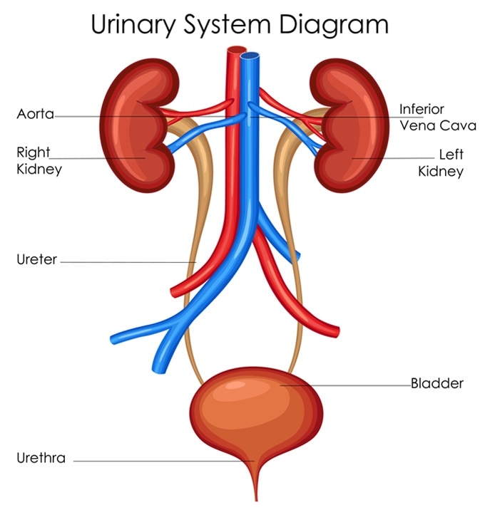
Like the stomach, the human bladder is a sac-like organ that expands and contracts when emptying. Located in the pelvis, above and behind the pubic bone, the bladder’s main function is to collect and store urine produced by the kidneys.
Features and Structure of the Bladder
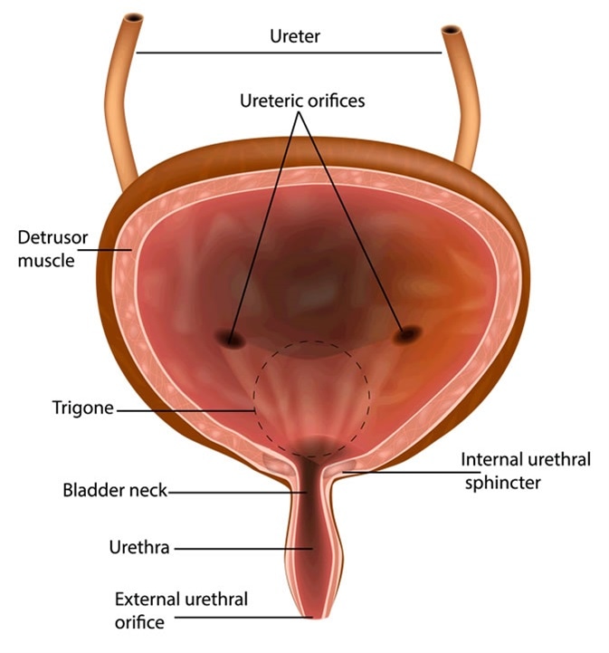
The transitional epithelium layer is the first layer on the inside of the –bladder. This acts as a lining that expands as the bladder fills.
The surrounding layer is the lamina propria, which is comprised of adipocytes, fibroblasts, nerve endings, and interstitial cells, which form an extracellular matrix. Encompassing this, is the muscularis propria layer (destrusor muscle) made up of thick, smooth muscle bundles.
The outermost layer is the perivesical soft tissue, made up of fat, fibrous tissue, and blood vessels. This layer separates the bladder from neighboring organs such as the kidneys and the prostate.

Ureter
The ureters are tubes which expel urine from the kidneys. Within the human body there are two ureters, one connected to each kidney.
The upper half of the structure is situated in the abdomen and the lower half in the pelvic area.
In the average adult, the ureter is 10 to 12 inches long. The tubes are able to contract due to fibrous and muscular walls coated in mucus.
Urethral Sphincter
There are two types of urethral sphincters: the internal urethral sphincter (IUS), characterized by smooth muscle and is under involuntary control, and the external urethral sphincter (EUS), where control is voluntary and consists of striated muscle.
Both structures are essential for urinary continence. During urination, the destrusor muscles of the bladder contract, and the sphincters relax and open in an antagonistic style, to allow the urine to exit the bladder into the urethra and out of the body.
When comparing the structure in men and women, there are a few notable differences. In women, the EUS tends to be more intricate compared to in men. Furthermore, the muscles in the EUS in women are involved in the constriction of the urethra and vagina.
Trigone
The trigone is a triangular muscular structure located between the bladder and urethra. Effective connection between the ureters and the trigone are vital for proper functioning of the ureteral valve mechanism.
The mechanism prevents backflow of urine from the bladder to the ureters which can cause damage to the kidneys and lead to end-stage renal disease.

Structure of the Bladder when Expanding
When the ureters remove urine from the kidneys to the bladder and the bladder begins to fill, the bladder’s muscles wall begin to thin, moving the bladder upwards and towards the abdominal cavity.
In contrast, when the bladder is empty, the muscle wall is thicker and the bladder as a whole becomes firmer.
The ability of the bladder to expand enables its size to increase from approximately two inches to over six inches long. For most individuals, their bladder reaches its upper capacity when storing between 400-600 ml.
Malfunction of the Bladder and its Features
If the urethral sphincter malfunctions it can cause lower urinary tract disorders. One common disorder is urinary incontinence (UI) and is characterized by the involuntary loss of urine.
The disorder affects both sexes, but is nearly twice as prevalent in women than in men. In women, vaginal childbirth can cause damage to ligaments, the pelvic floor and facial support, ultimately causing UI.
Individuals can also experience an increase in the need to urinate more frequently, which can be caused by a reduced capacity of the bladder.
Cystitis is another common bladder condition marked by inflammation of the bladder which typically results from a bladder infection. Most cases are mild, and will often improve without any medicinal intervention. However, for some, recurrent bouts of cystitis may require long-term treatment, and in extreme cases could cause kidney infection.
The bladder is comprised of, and works with, several other structures within the human body. Promoting good bladder health is essential for preventing the onset of malfunction, which can have varying consequences such as urinary incontinence, cystitis and the development of cancer.
Sources
- Andersson, K. E., & McCloskey, K. D. (2014). Lamina propria: the functional center of the bladder?. Neurourology and urodynamics, 33(1), 9-16. DOI: https://doi.org/10.1002/nau.22465
- Bladder: https://www.healthline.com/human-body-maps/bladder#3
- Jung, J., Ahn, H. K., & Huh, Y. (2012). Clinical and functional anatomy of the urethral sphincter. International neurourology journal, 16(3), 102. DOI: 10.5213/inj.2012.16.3.102
- Picture of the Bladder: www.webmd.com/urinary-incontinence-oab/picture-of-the-bladder#1
- Ureter: https://www.healthline.com/human-body-maps/ureter#1
- Viana, R., Batourina, E., Huang, H., Dressler, G. R., Kobayashi, A., Behringer, R. R., … & Mendelsohn, C. (2007). The development of the bladder trigone, the center of the anti-reflux mechanism. Development, 134(20), 3763-3769. DOI:10.1242/dev.011270
Further Reading
- All Bladder Content
- How to Keep a Bladder Diary
- Bladder Control and Diet Changes
Last Updated: Sep 2, 2018
Source: Read Full Article
