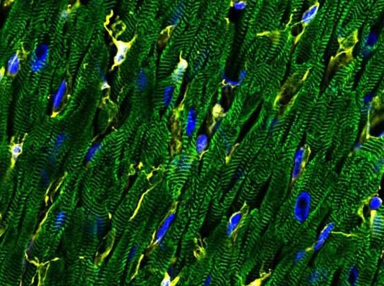
Single-cell map of the heart reveals wide cellular diversity

The largest single-nucleus survey of the human heart to date uncovers several new cell types and a variety of gene expression patterns.
The study of individual cells has vastly expanded the biological understanding of various organ systems, but in cardiac research, some basic questions have remained. How many types of cells make up the human heart? Do these cells behave differently in different regions of the heart? How do various cell types influence heart health and disease?
To help answer these questions, researchers from the Precision Cardiology Lab (PCL) of the Broad Institute of MIT and Harvard and Bayer, Massachusetts General Hospital, University of Pennsylvania, and Masonic Medical Research Institute have now generated the most comprehensive high-resolution cellular map to date of the human heart. By studying nearly 300,000 individual heart cells from seven healthy hearts, the team identified nine major cell types and over 20 subtypes in the organ. They also found gene expression patterns across different parts of the organ, as well as cell types that may be involved in common cardiovascular diseases.
Understanding all the distinct cells—and how they vary—within the healthy human heart allows researchers to home in on how these cells behave differently in disease. This knowledge could help scientists identify novel drug targets in specific cardiac cell types implicated in cardiovascular disorders. This work appears in Circulation.
“Not a lot was known about the different types of cells in the human heart,” said Christian Stegmann, the Bayer head of the PCL.
Senior study author Patrick Ellinor agreed. “At a fundamental level, we were trying to ask something really simple, which is how many cell types are in the human heart, and what do they look like,” said Ellinor, a Broad institute member, director of the PCL and the Broad’s Cardiovascular Disease Initiative, and director of the Cardiac Arrhythmia Service at Massachusetts General Hospital.
Heart of the cells
Decades of cardiac research have focused on the heart’s muscular component, with cells known as cardiomyocytes, that helps the heart contract. But cardiomyocytes rely on an intricate ensemble of specialized cells to keep hearts pumping, and researchers have long debated what the major cell types within the human heart are.
“In recent years, we have come to appreciate that the heart is a really complex mixture of cells,” said Nathan Tucker, first author of the study and a project team lead in the PCL, who is now an assistant professor at the Masonic Medical Research Institute. “There’s a lot of untapped biology for us to understand.”
Human heart cells have been challenging to study, even with modern single-cell sequencing techniques, because cardiomyocytes are too large to be processed by the standard fluidic devices used in these methods. Instead, the team behind this new study adopted a different approach: single-nucleus sequencing, which breaks up the cells and analyzes the RNA in individual nuclei rather than from whole intact cells. This approach allowed them to survey cells across the human heart, define their gene expression profiles, and probe rarer cell types beyond what was previously possible.
The team identified the surprising number of heart cell types and subtypes by profiling 287,269 individual nuclei from all four chambers of the heart. “Many people think of the heart as relatively homogeneous,” Ellinor noted. “The number of cell subtypes, the extent of the diversity, and just the sheer complexity were humbling.”
The single-nucleus data also showed that cellular gene expression profiles varied by heart chamber in certain cell types. For example, the heart’s resident immune cells displayed a distinct gene expression pattern in the right atrium as compared to the other chambers.
“We’ve long understood that cardiomyocytes across the heart’s chambers are different. But we didn’t know that all the other support cells and their activity would also be different across the different chambers,” said Tucker. “So, the fact that you need to understand these cells in the context of the chamber that they come from was really a surprising and important finding.”
Total map of the heart
The study also creates a critical new resource for the scientific community: a reference map of the heart to better understand what goes awry in disease.
Cardiovascular diseases, such as heart failure and stroke, kill millions around the world every year. Understanding the genetics of these diseases has been challenging without a baseline cellular map of a healthy human heart.
To help put heart disease genetics into a cellular context, the researchers coupled their single-nucleus data with genetic association data from previous population-based disease studies. They identified cell types within the heart that are most likely to have a role in common cardiovascular diseases such as heart attack, and atrial fibrillation, where the heart beats irregularly.
The scientists also looked for cardiac cell types that express genes with known links to disease, to identify potential new drug targets for cardiovascular disease. They found some striking patterns. Most of the druggable genes they explored were expressed in just three cardiac cell types: fibroblasts cells that manufacture and maintain connective tissue, cardiomyocytes, and fat cells.
Collectively, these insights into the heart’s individual cells and how they vary in health and disease will enable researchers to identify new therapeutic targets in disease-specific cell types, powering drug discovery efforts for cardiovascular diseases.
In fact, the research team will soon begin studying individual cells from patients with certain cardiac diseases to learn how various types of heart cells behave in a diseased condition.
Source: Read Full Article
