Applications of CLARITY
CLARITY or Clear Lipid-exchanged Acrylamidehybridized Rigid Imaging/Immunostaining/In situ-hybridization-compatible Tissue-hYdrogel is a method of histological fixation and clearing.
CLARITY is used to perform immunohistochemistry as it maintains fluorophores while imaging and performing 3D reconstruction for large volumes. This method is based on creating a hydrogel that removes the lipids and keeps the proteins and DNA of tissue intact.
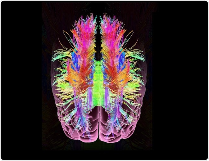
 Michael Braham | Shutterstock
Mechanism of CLARITY
CLARITY involves creating a tissue-hydrogel hybrid by polymerizing proteins along with DNA and RNA using acrylamide. This is based on the ability of acrylamide to bind molecules that have amine ends, including proteins, DNA, and RNA. This creates a skeletal structure that larger proteins can still permeate.
Detergent is then added to encapsulate lipids into micelles that are then carried through the pores of the skeletal structure using an applied electrical field. The antibodies to be used in an immunoassay are subsequently added and they can also pass through the pores and bind to membrane proteins.
The step-by-step procedure for CLARITY creation is: fixing the tissue, forming hydrogels, extracting the lipids, and matching the refractive index.
Imaging Human Samples
One potent application of CLARITY lies in its ability to allow clear imaging of the human brain during pathogenesis or diseased state. The cases that have been visualized using CLARITY include Alzheimer’s, Parkinson’s, neurodegeneration during mitochondrial disease, autism, and intractable epilepsy. These were visualized in tissues that were fixed using formalin.
Brain samples that have been fixed in paraformaldehyde and subjected to cryopreservation in sucrose have also been examined using CLARITY. In these cases, myelination and duration of fixation in formalin often determines the speed with which tissue clearance is obtained. However, in some cases, the tissue has been observed to expand transiently when the tissues were exposed to tissue clearing methods for extended durations, such as greater than 40 days.
Imaging the Nervous System and Vasculature
Studies show that skeletal muscle can be cleared using the technique of CLARITY; however, it is more difficult to clear neuromuscular junctions. This is because the extensive cross-linking and fixation at these junctions often prevent the access of labeled probes to the acetylcholine receptors.
It has, however, been possible to target the peripheral nerves using immunohistochemistry or lipophilic tracers. Endothelial cells can also be imaged by treatment with a Dil-dye or biocytin before fixing.
Imaging Muscle and Bone
Tendons are rich in collagen making them opaque and difficult to clear. This can be improved by increasing the clearing time during CLARITY and also adjusting the refractive index. Chondrocytes or cells that make cartilage can be stained using lipophilic dye, such as Dil-SP.
Bone tissue has low lipid content, additional mineral content, and bone marrow. These tissues can be cleared using solvent-based methods or by adding a decalcification step. In this step, the tissue is kept intermittently in EDTA and SDS at increased pH.
This modification generates a visualization depth of 200–300µm. The calcium removal step can provide an additional visualization depth of 1.5 mm. The bone marrow is also rich in heme protein that generates autofluorescence. This is adjusted by removing the heme moiety using amino alcohol before the process of refractive matching.
Imaging other Organs and Species
CLARITY has been used to image several organs, such as the liver, kidney, pancreas, adrenal glands, spleen, lymphoid tissue, intestine, testes, ovaries, lung tissue, etc. However, the protocol must be modified based on the tissue type as different tissues have different densities, ratios of proteins and lipids, clearing times, and the average refractive index of the hydrogel.
This method has also been performed on other organisms, such as rabbits, chickens, zebrafish, Xenopus, octopus, and others. In the case of plants, an additional degradation step is added to remove the cell wall post tissue clearing. This is done to clear and stain the cells without resorting to sectioning of the tissue.
Sources
- https://www.ncbi.nlm.nih.gov/pubmed/28728966
- www.biorxiv.org/content/biorxiv/early/2017/06/02/144378.full.pdf
- www.ncbi.nlm.nih.gov/pmc/articles/PMC4092167/pdf/nihms596795.pdf
Further Reading
- All CLARITY Content
- What is the CLARITY Technique?
- CLARITY Methodology
Last Updated: Apr 5, 2019

Written by
Dr. Surat P
Dr. Surat graduated with a Ph.D. in Cell Biology and Mechanobiology from the Tata Institute of Fundamental Research (Mumbai, India) in 2016. Prior to her Ph.D., Surat studied for a Bachelor of Science (B.Sc.) degree in Zoology, during which she was the recipient of anIndian Academy of SciencesSummer Fellowship to study the proteins involved in AIDs. She produces feature articles on a wide range of topics, such as medical ethics, data manipulation, pseudoscience and superstition, education, and human evolution. She is passionate about science communication and writes articles covering all areas of the life sciences.
Source: Read Full Article
