Towards better dental cavity diagnosis with nondestructive infrared imaging

Dental caries, also called “cavities” or “tooth decay,” can be debilitating. Modern dentistry has tried-and-tested solutions for treating caries of many shapes and sizes without needing to remove the tooth altogether. Dental restorations, also known as “fillings,” are commonplace and yet—while modern restorative materials have many advantages such as improved biocompatibility and better esthetics—they can still fail over time.
Restorative materials do not always bond well to the surrounding healthy tooth structure. Microscopic leaks may form, allowing fluids and bacterial acids to penetrate beneath the restoration. This can lead to the formation of a secondary caries that appears and grows around a previously restored cavity.
“Dentists now spend more time replacing failed restorations than placing new ones due to the maladaptation of bonding materials to tooth structure,” comments Dr. Nai-Yuan N. Chang from the Fried Group in the Department of Preventive and Restorative Dental Sciences at University of California, San Francisco (UCSF).
Directed by UCSF Professor Daniel Fried, the Fried Group develops new diagnostic techniques to detect active dental lesions. In a recent study published in Journal of Biomedical Optics (JBO), Chang’s research team evaluated emerging imaging modalities for discerning active tooth decay.
“The traditional methods relying on tactile sensation via a dental explorer and visual inspection based on texture and color are highly subjective and unreliable,” explains Chang. “However, there is currently no established dental imaging technology that can provide diagnostic information with high specificity and sensitivity when assessing dental decay activity.”
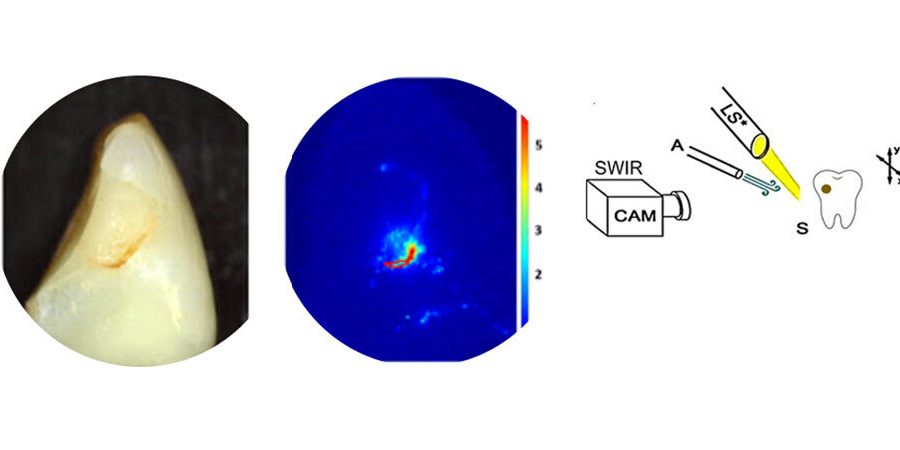
To address this issue, the researchers focused on whether shortwave-infrared (SWIR) and thermal imaging could be combined with air drying to accurately diagnose the activity of a secondary caries lesion. The idea underlying both these methods is that active lesions are more porous than healthy tooth, and these pores hold water.
In the SWIR-based approach, one can indirectly detect active lesions by observing changes in the SWIR reflectivity as the tooth dries out. On the other hand, the thermal imaging-based approach relies on the fact that the temperature changes in active lesions during air drying are different from that in healthy tooth, owing to the water trapped in the pores of the lesion.
In their work, the team acquired 63 human tooth samples from oral surgeons and analyzed 109 suspected secondary lesions in them using both SWIR and thermal imaging. In addition to these methods, the researchers also observed the samples using optical coherence tomography (OCT), a more sophisticated technique that uses near-infrared light to create high resolution 3-D images.
To determine whether SWIR and thermal imaging were indeed useful for detecting active lesions, the results of these methods were compared with those obtained via OCT.
Overall, SWIR proved superior to thermal imaging and performed better in most circumstances. The SWIR permeability measurements were well correlated with the thickness of the transparent surface layer (TSL) of lesions measured via OCT.
The team found that the highly mineralized TSL was thickest when a lesion had been fully arrested and needed no further intervention. According to the OCT results, a TSL thicker than 70 µm was a potential indication that a lesion was no longer active.
The findings of this study could help pave the way to a new era of diagnostic imaging in dentistry. Excited about the results, Chang concludes, “Our work provides further developmental milestones towards meeting the need for better diagnostic and easily operable clinical devices.”
More information:
Nai-Yuan N. Chang et al, Assessment of the activity of secondary caries lesions with short-wavelength infrared, thermal, and optical coherence tomographic imaging, Journal of Biomedical Optics (2023). DOI: 10.1117/1.JBO.28.9.094801
Journal information:
Journal of Biomedical Optics
Source: Read Full Article