Managing variety in MRI scans can lead to better stroke diagnoses
 for his Master’s project. Credit: © 2022 EPFL / Alain Herzog")
The first few hours following a stroke are crucial. To be able to treat a patient effectively, doctors must rapidly localize the damaged blood vessel and determine what kind of stroke occurred. In most cases, either a ruptured blood vessel releases blood into the brain, or a blood clot obstructs a blood vessel in the brain. Patients who experience the second type of stroke are prescribed medication to dissolve the blood clot. If this medication is given to patients of the first type, however, it will fluidify the blood and only make the hemorrhaging worse.
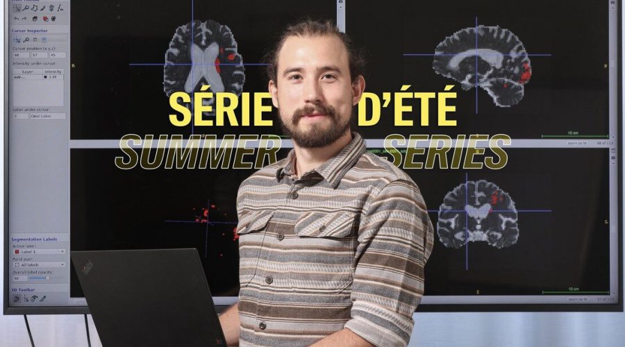
Yet doctors must take action quickly because the faster a stroke is treated, the lower the likelihood of severe consequences. “Stroke patients are given an MRI [magnetic resonance imaging] scan immediately upon arriving at the hospital,” says Antoine Madrona, who is completing a Master’s degree in life sciences. “The scan is used to confirm that it’s indeed a stroke and to identify what type, in order to prescribe the right treatment.”
Applying AI to medical imaging
Deep-learning algorithms that sort through vast data sets to generate predictions can help doctors make the right diagnosis. They can show radiologists where they should focus their attention, provide quantitative information on the position, size and number of blood vessel injuries, and speed the process of selecting a treatment. As part of his Master’s project, Madrona helped develop an algorithm based on data from diffusion-weighted magnetic resonance imaging (DW-MRI)—a form of MRI that uses the diffusion of water molecules to generate contrast in images.
The DW-MRI data sets that Madrona obtained from various Swiss hospitals were extremely heterogeneous. “I was surprised to see so much variation in the images from different hospitals,” he says. “I didn’t expect to see such little standardization.”
In other words, the same patient undergoing an MRI in two different hospitals will end up with two very different images. That’s due to variations in the machine model and the image acquisition protocol. “There’s no national or even international standard,” says Madrona. “Each hospital uses its own magnetic pulse sequence and sets its own pulse directions and intensities. All that affects the image contrast and overall appearance.”
Data security
To make sure patients’ data remain confidential, Madrona used a federated learning method for his algorithm. This method entails training an algorithm across several data sets but without exchanging any data between them. None of the medical imaging systems currently on the market employs a federated method. “I’m particularly interested in how decentralized algorithms can be used to protect patient data,” he says. “This gets to an important ethical issue that I believe needs to be addressed more widely in the health care industry. Patient confidentiality shouldn’t be the price to pay for more efficient diagnostics.” Madrona, now 25, intends to pursue a career in this direction.
Source: Read Full Article